
Your eye doctor can spot potential health issues beyond just vision problems. In fact, with the help of retinal imaging, we can look deeper into your overall health. Retinal imaging involves taking highly detailed pictures of your retina—the light-sensitive layer at the back of your eye. These images can reveal early signs of eye diseases like glaucoma or macular degeneration, systemic conditions like diabetes or high blood pressure, and important defects like holes or tears.
Retinal imaging is like a window into your health, helping your doctor catch problems early and keep you feeling your best.
Why Is the Retina Important?
The retina acts as the gateway to your vision. It’s an intricate layer of tissue lining the back of your eye that transforms rays of light into electrical signals your brain then interprets as images. Without a functioning retina, the brain can’t process visual information.
Beyond its role in creating vision, the retina can also serve as a window into your overall health. Changes in its structure can reveal underlying systemic issues, such as diabetes, high blood pressure, or autoimmune diseases, making it important for medical diagnostics. We must also view this important liner in order to make sure there are no compromises starting, such as thinning, holes or tears present.
This is why imaging the retina is such a key aspect of modern eye care.
Understanding Retinal Imaging: A Closer Look
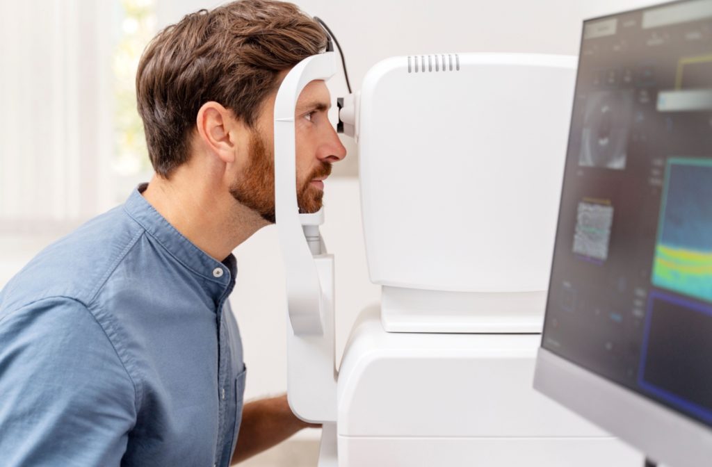
Retinal imaging is a fast, non-invasive procedure that captures detailed pictures of your retina. It allows the doctor to get a clear view of your eye’s interior structure.
Several advanced techniques make retinal imaging an essential diagnostic tool:
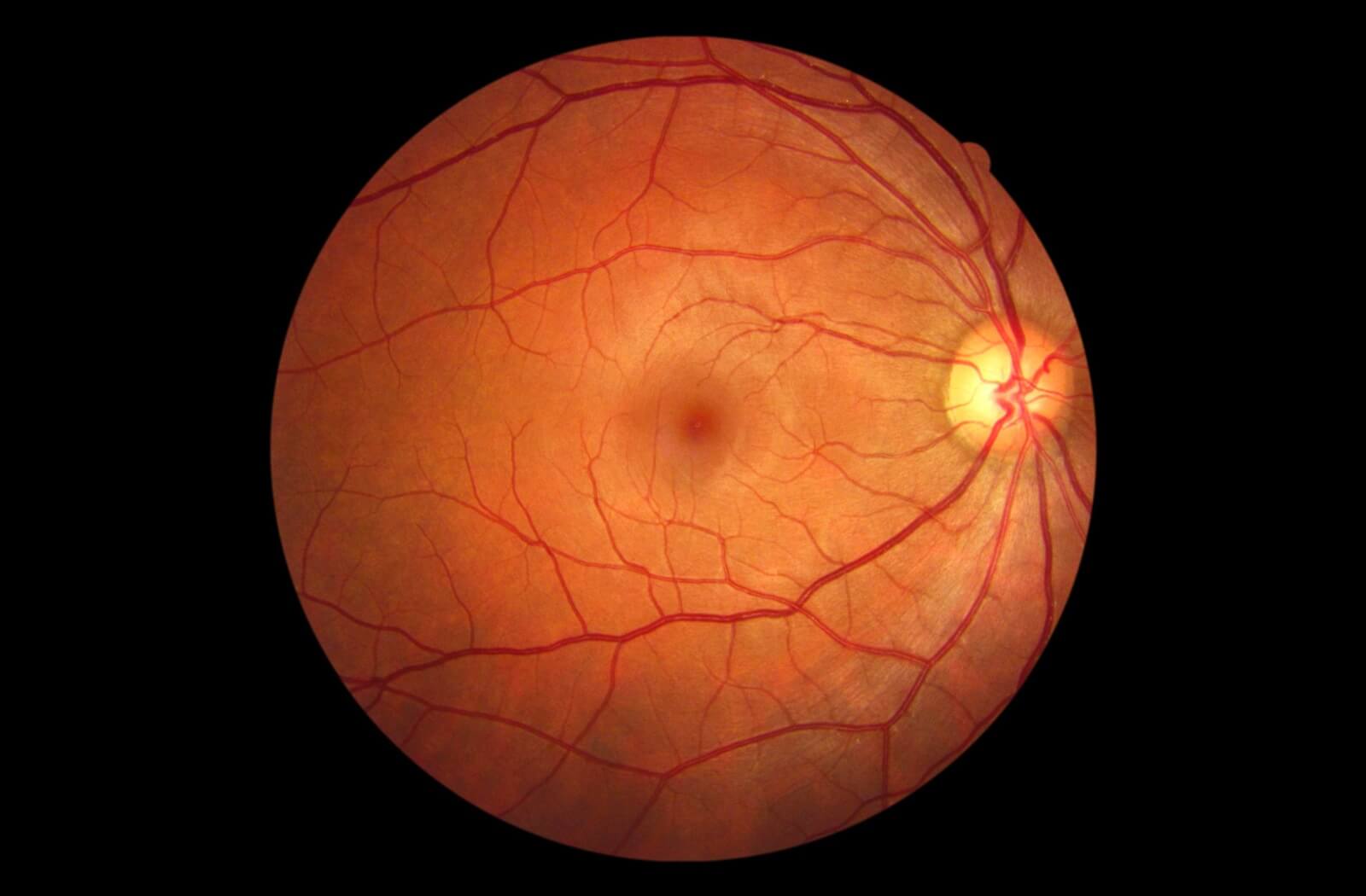
Fundus Photography
This is one of the most common methods for imaging the retina. It uses a specialized camera to take high-resolution colour images of the retinal surface, displaying up to 50 degrees of the central retina. Fundus photographs reveal early signs of conditions like diabetic retinopathy, hypertensive retinopathy, optic nerve conditions and macular degeneration.
Optical Coherence Tomography (OCT)
OCT uses light waves to create cross-sectional images of the retina, much like a 3D scan. It’s particularly helpful in diagnosing conditions like glaucoma or macular edema by highlighting changes in the layers of the retina.
Fluorescein Angiography
Fluorescein angiography uses a fluorescent dye injected into your bloodstream to assess the blood flow in your retina. A special camera then tracks the dye as it moves through the blood vessels, providing insights into conditions like diabetic retinopathy and wet macular degeneration.
The Significance of Ultra-Widefield (UWF) Imaging
While traditional fundus photography captures important central retina details, it will miss issues in the peripheral areas of the retina. That’s where ultra-widefield imaging (UWF) shines.
UWF imaging technology, such as the Optos optomap, provides a 200° view of your retina in a single image—covering over 80% of the retinal surface. This far exceeds the capabilities of conventional imaging systems, which typically capture only 15-30% of the retina at a time.
The benefits of UWF include:
- Detecting issues early: UWF imaging can detect peripheral retinal problems, such as tears or detachments, that might otherwise go unnoticed.
- Comprehensive view before exams: With UWF technology, doctors can assess the majority of the retina even before other scans or tests, enabling quicker and better-informed decisions.
- Time-efficient: It enhances practice efficiency by reducing the time spent on detailed examinations, without sacrificing diagnostic precision.
For these reasons, many clinics have begun incorporating UWF imaging as a routine part of eye exams.
What Can Retinal Imaging Detect?
Retinal imaging is an effective diagnostic tool that’s capable of identifying a wide range of eye conditions. Here’s a closer look at some of the conditions it can help detect:
- Diabetic Retinopathy: Detects signs of this diabetes-related eye condition, including abnormal blood vessels or bleeding in the retina, often before symptoms appear.
- Age-Related Macular Degeneration (AMD): Identifies damage to the macula, which can lead to central vision loss in older adults.
- Glaucoma: Highlights changes in the optic nerve and retinal nerve fiber layer, which can indicate this pressure-induced eye disease.
- Retinal Detachment: Reveals holes, tears or full detachment of the retina—issues that require immediate surgical intervention.
The retina is fairly unique in how much information it can give us about the body as a whole. Because of this, retinal imaging can also reveal systemic health issues that may typically slip under the radar, including:
- Hypertension: Tracks changes in retinal blood vessels resulting from high blood pressure, such as narrowing or leakage.
- Multiple Sclerosis (MS): Detects alterations in the optic nerve that may be indicative of MS.
- Autoimmune Diseases & Other Factors: Pinpoints inflammation, infections, or signs of broader health issues reflected in the eye.
Retinal imaging isn’t just about protecting your vision; it’s also a stepping stone toward better overall health management.
See the Bigger Picture with Retinal Imaging
Your eyes are more than windows to your soul—they’re windows to your health. Retinal imaging combines advanced technology and medical expertise to bring you clearer insights into both your eye and systemic health.
If you’re due for an eye exam, don’t wait to take control of your eye health. Book an appointment with Discover Eyecare. We leverage the power of retinal imaging technology like Ultra Wide Field Imaging and OCT, alongside other diagnostic technologies and compassionate care to give you the vision you deserve.
Experience a clearer view of your health—schedule your visit with us today!